Part I of this series includes discussions on protocol, anatomy and a case review session centered around ocular pathology. Along the way Dr. Yousem shares the pearls and pitfalls of imaging this area, garnered over years of experience and interest in this topic. This series is eligible for 2 AMA PRA Category 1 CME credits.
In the second installment of the orbit course, we dig deep into the case review for abnormalities of the intraconal space. This segment adds 15 new videos and a dozen case reviews to help round out your knowledge of orbit pathology. This series is eligible for 1.25 AMA PRA Category 1 CME credits.
The final section focuses on conal and extraconal pathology. The video lectures discuss the conal and extraconal spaces as well as the associated appendages to examine disease processes and imaging appearances. This series of lectures completes our look at some of the common pathologies that affect the orbits. This series is eligible for 1.5 AMA PRA Category 1 CME credits.
Some of the diagnoses included in this course:
- Endophthalmitis
- Juvenile Ossifying Fibroma
- Lacrimal Gland Lymphoma
- Microphthalmia
- Neuromyelitis Optica
- Neuromyelitis Optica Spectrum Disorder
- Optic Neuritis
- Orbital Fractures
- Orbital Pseudotumor
- Periorbital Cellulitis
- PHPV
- Pilocytic Astrocytoma
- Retinal detachment
- Retinoblastoma
- Staphyloma
- Thyroid Eye Disease
- Venous Vascular Malformation
- And more…
Objectives :
After completing this course, you will be better able to:
- Apply appropriate search patterns to ensure high quality case assessment
- Identify key anatomical landmarks, variations, and abnormalities on imaging
- Accurately interpret advanced imaging cases
- Formulate definitive diagnoses and limited differentials
Topics:
Introduction to Imaging of the Orbits
Orbit Protocols for CT
Orbit Protocols on MRI
Imaging Artifacts in MRI and CT
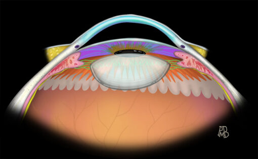
Anterior Globe Anatomy
Membranes of the Orbit
Anatomic Considerations: Orbital Spaces
The Conal Space
Extraconal Space
Orbital Boundaries
Orbital Appendages
Orbit Anatomy on CT
Axial Anatomy on MRI
Coronal Anatomy on MRI
Anterior Globe Rupture with Laterally Dislocated Cataract
Foreign Body in Globe
Wood Foreign Body and Ocular Hypotony
Hemmorhage in Both Chambers, Open Globe
Staphyloma
Microphthalmia, PHPV
Retinal Detachment
Retinoblastoma
Retinoblastoma on MRI
Retinoblastoma in a Pediatric Patient
Ocular Pathology – Review – 10 min
Endophthalmitis
PHPV Review
Phthisis Bulbi
Ocular Calcification
Retinoblastoma – Review
Choroidal Melanoma
Intraconal, Conal and Extraconal Anatomy
Intraconal Hemangioma
Venous Vascular Malformation
Optic Nerve Glioma, NF1
Pilocytic Astrocytoma
Multiple Sclerosis, Optic Neuritis
Neuromyelitis Optica
Neuromyelitis Optica Spectrum Disorder
Neuromyelitis Optica With Spinal Cord Involvement
Optic Nerve Sheath Meningioma
Bilateral Optic Neuritis, Leukemia
Intraconal Pathology – Review
Optic Neuritis – Review
Optic Nerve Glioma – Review
Optic Nerve Sheath Meningioma – Review
Introduction to Conal Lesions
Lipogenic Thyroid Eye Disease
Thyroid Eye Disease
Orbital Pseudotumor
Conal Pathology – Review
Anatomic Review – Extraconal Spaces
Periorbital Cellulitis & Abscess
Type 3 Orbital Infection
Solitary Fibrous Tumor
Langerhans Cell Histiocytosis
Juvenile Ossifying Fibroma
Perineural Spread of Squamous Cell Carcinoma
Diffuse Metastatic Disease to Bone with Extraosseous Extension to the Extraconal Space
Orbital Floor Fracture
Orbital Floor Fracture with Muscle/Fat Herniation
Orbital Floor Fracture: Status Post Repair
Bilateral Orbital Fracture Repair
Review – Periorbital Cellulitis
Orbital Pseudotumor – Review
Orbital Wall Abnormalities – Review
Orbital Fracture – Review
(Extraconal) Giant Cell Reparative Granuloma
(Extraconal) Granulomatis Sinusitis Associated with IgG4-related Ophthalmic Disease
Anatomic Review – Orbital Appendages
Squamous Cell Carcinoma of the Nasolacrimal Sac
Sarcoidosis of the Lacrimal Gland
Lymphoma of the Lacrimal Gland
Adenoid Cystic Carcinoma of the Lacrimal Gland
Orbital Appendage Pathology – Review
Content reviewed: August 22, 2019


