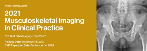
This activity provides a review of clinical applications concerning the diagnosis, treatment and management of the musculoskeletal disorders. Modern imaging strategies, surgical correlation and the need for intra-disciplinary teamwork when deciding the most effective patient management is addressed. Faculty share techniques and tips in image interpretation of musculoskeletal injuries and pathology. Information gained from this activity is geared to improve diagnostic capabilities and expand clinical applications.
Target Audience
This CME activity is designed to educate diagnostic imaging physicians who supervise and interpret musculoskeletal MRI. In addition, referring physicians who order musculoskeletal MRI should gain an appreciation of the strengths and limitations of these types of procedures.
Educational Objectives
At the completion of this CME teaching activity, you should be able to:
- Optimize image protocols to accurately assess musculoskeletal injury and pathology.
- Assess patients with joint pathology in a non-invasive manner utilizing MRI.
- Recognize the strengths and limitations of MRI for the management of sports related injuries.
- Describe the MR appearance of muscle and tendon injury.
- Differentiate musculoskeletal masses and tumors.
- Correlate MRI, ultrasound and surgical findings of musculoskeletal injury.
Program
Musculoskeletal Disease: Unifying Concepts
William B. Morrison, M.D., FACR
How to Avoid Your Case Being Presented at Next M&M
Tetyana Gorbachova, M.D.
Imaging Pearls I Would Like to Tell My Younger Self
Mark Cresswell, MBBCh, BSc (Hon), FRCR, FRCPC
Orthopedic Interposition Injuries: You Only See What You Look For
Robert D. Boutin, M.D.
Radiographic / MRI Correlation
William B. Morrison, M.D., FACR
MSK Hot Topics
Robert D. Boutin, M.D.
Optimization of MSK MRI
William B. Morrison, M.D., FACR
MRI of the High Performance Athlete
Adam C. Zoga, M.D., M.B.A.
MRI of the Throwing Athlete’s Shoulder
Lawrence M. White, M.D., FRCPC
Sports-specific Injuries
Adam C. Zoga, M.D., M.B.A.
Trauma of the Knee
Tetyana Gorbachova, M.D.
MRI of the Knee: Case-Based Review – Misses That Matter
Robert D. Boutin, M.D.
MRI of the Knee: Cruciate Anatomy and Injury Patterns
William B. Morrison, M.D., FACR
MRI of the Knee Menisci
Tetyana Gorbachova, M.D.
Knee MRI: Post-op Case-Based Review
Robert D. Boutin, M.D.
Shoulder Impingement and the Rotator Cuff
Adam C. Zoga, M.D., M.B.A.
MRI of Shoulder Instability
Lawrence M. White, M.D., FRCPC
Imaging of the Brachial Plexus
Mark Cresswell, MBBCh, BSc (Hon), FRCR, FRCPC
MRI of the Elbow
Tetyana Gorbachova, M.D.
Elbow: MRI Case-Based Review
Robert D. Boutin, M.D.
Wrist Imaging: Location, Location, Location
Mark Cresswell, MBBCh, BSc (Hon), FRCR, FRCPC
MRI of the Wrist and Hand
Tetyana Gorbachova, M.D.
Ankle MRI: The Essentials
William B. Morrison, M.D., FACR
Ultrasound / MRI Correlation
Mark Cresswell, MBBCh, BSc (Hon), FRCR, FRCPC
Bone Tumors: Principles of Diagnosis and Treatment
John A. Abraham, M.D., FACS
0.25 Hrs
Imaging of Neural Impingement
Mark Cresswell, MBBCh, BSc (Hon), FRCR, FRCPC
Soft Tissue Tumors: Principles of Diagnosis and Treatment
John A. Abraham, M.D., FACS
Post-op MRI/Surgical Correlation: What the Surgeon Wants to Know
John A. Abraham, M.D., FACS
MRI of Bone Marrow
Tetyana Gorbachova, M.D.
MRI Evaluation of Total Hip Arthroplasty
Lawrence M. White, M.D., FRCPC
Athletic Pubalgia and Core Injury
Adam C. Zoga, M.D.
CAM Femoroacetabular Impingement: What Is It and How Should We Assess It?
Lawrence M. White, M.D., FRCPC
Hip: Periarticular Pathology
Adam C. Zoga, M.D., M.B.A.
Spine MRI: MSK Perspective
William B. Morrison, M.D., FACR
CME Release Date 9/15/2021
CME Expiration Date 9/14/2024


